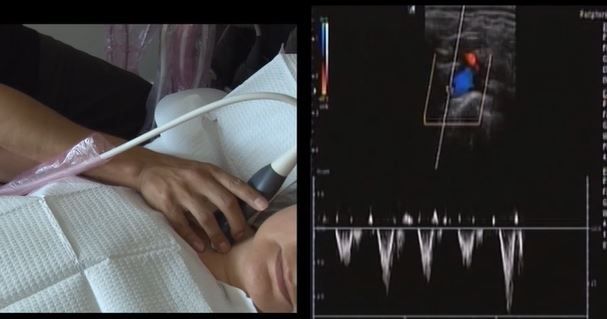
Upper extremity arterial ultrasound
-
26 minutes
Video duration

Learning objectives
- review about the anatomy of the upper extremity arteries using props on a real patient
- learn about scanning techniques to image the upper extremity arteries using ultrasound
- understand about transducer manipulation and image optimization applicable to imaging the upper extremity arteries using ultrasound
- simultaneously view transducer manipulation, window selection, and the resulting ultrasound image
- become familiarized with an example of a protocol to image the upper extremity arteries using ultrasound, including one to rule out thoracic outlet syndrome
What's included in this online course?
A step-by-step guide
Study at your own pace
Take notes as you watch and get prepared to practice in a lab setting.
Unique learning experience
Step-by-step explanation of scanning techniques by Leonardo while scanning.
Summary of important points.
Leonardo Faundez
MA-ED BSc DMS RDMS RVT CRGS CRVS
Leonardo graduated from the Ultrasound Technology Program from The Michener Institute of Education at UHN in 1997. In 2008 he completed a master’s degree in education at Central Michigan University.
Leonardo has been in the ultrasound field for 27 years as a registered
Canadian diagnostic medical sonographer, educator, entrepreneur, and lecturer. He
worked as a full-time sonographer and clinical instructor for about 9 years at
University Health Network. He then went on to work as an Ultrasound Professor at The Michener
Institute of Education at UHN for 10 years.
Leonardo has presented at several provincial, national, and international conferences on several sonography topics.
In 2013, Leonardo started Aprende Canada, where he offers sonography continuing education.

